
We looked at red blood cells and saw magnificent contours on the donut shaped object. We also looked at paper — complex intertwined fibers with bits of ink materials coloring parts of it.
Truly, a whole new world opened in front of me.
That was 20 years ago. I went on to study biology and did research in many different organisms. In addition to the shape, the physiology is even more fascinating.
Watching the first beat of heart cells we differentiated from embryonic stem cells gives you the euphoria of understanding how things work. It’s not just one cell twitching, but a whole group contracting and slowly relaxing before the next contraction. They are alive!
Who’s giving the initiation signal? How do they know to contract together? Is electricity propagating through the cells when that’s happening? It opens a whole dimension of imagination to how these cells are communicating with each other and working together in your body.
Today I study neuroscience on a fascinating little worm. When I look into the microscope, I can see individual neurons light up while the worms perform a particular turn. It’s a chain reaction happening inside of their body as the neurons control the behavior.
Perhaps one day we will read human thoughts just as we read the neuronal movement commands from of a worm.
I guess I’m still obsessed with mechanisms in the microscopic world.
1. The Earth Dwelling nematode worm Caenorhabditis elegans
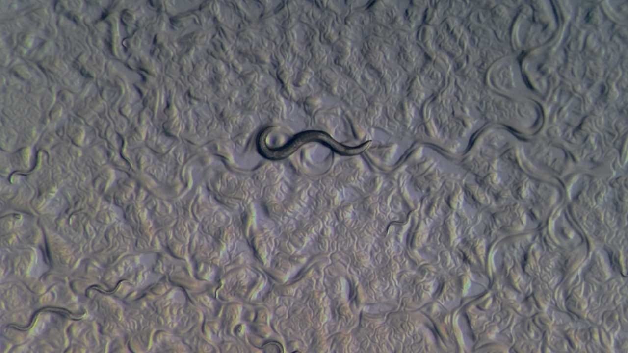
The Earth Dwelling nematode worm Caenorhabditis elegans.
The earth dwelling nematode worm C. elegans is model organism used by scientists to study the connections between neurons and behaviors. Their 302 neurons are readily seen through their transparent body. The little worm has very sharp sensations such as smelling odors that can’t be sensed by human, or temperature changes less than 0.1 degree.
Scene 1 shows the realtime speed of an adult C. elegans crawling on a phase contrast microscope with @iLabCam.
Scene 2 shows groups of C. elegans in a different stage of their life in time lapse movie under a dissecting microscope with LabCam.
Scene 3 shows a transgenic C. elegans under a fluorescent microscope with LabCam.
2. Western black-legged tick Ixodes pacificus after a big blood meal
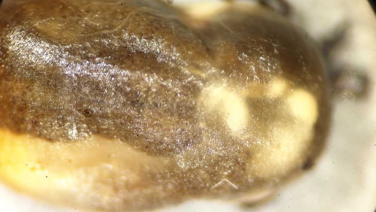
Western black-legged tick Ixodes pacificus after a big blood meal
You can see its intestines moving under its semi-transparent body shell with a dissecting microscope with LabCam. They can latch onto an animal for up to four days. They slowly expand from a small tick to a huge one while sucking blood. In Humboldt State University we studied the interaction between ticks and the bacteria that live in them, with the hope of finding a way of controlling tick-borne diseases. Credit: Du Cheng, Dr. Jianmin Zhong
3. A dish of beating heart cells differentiated from embryonic stem cells
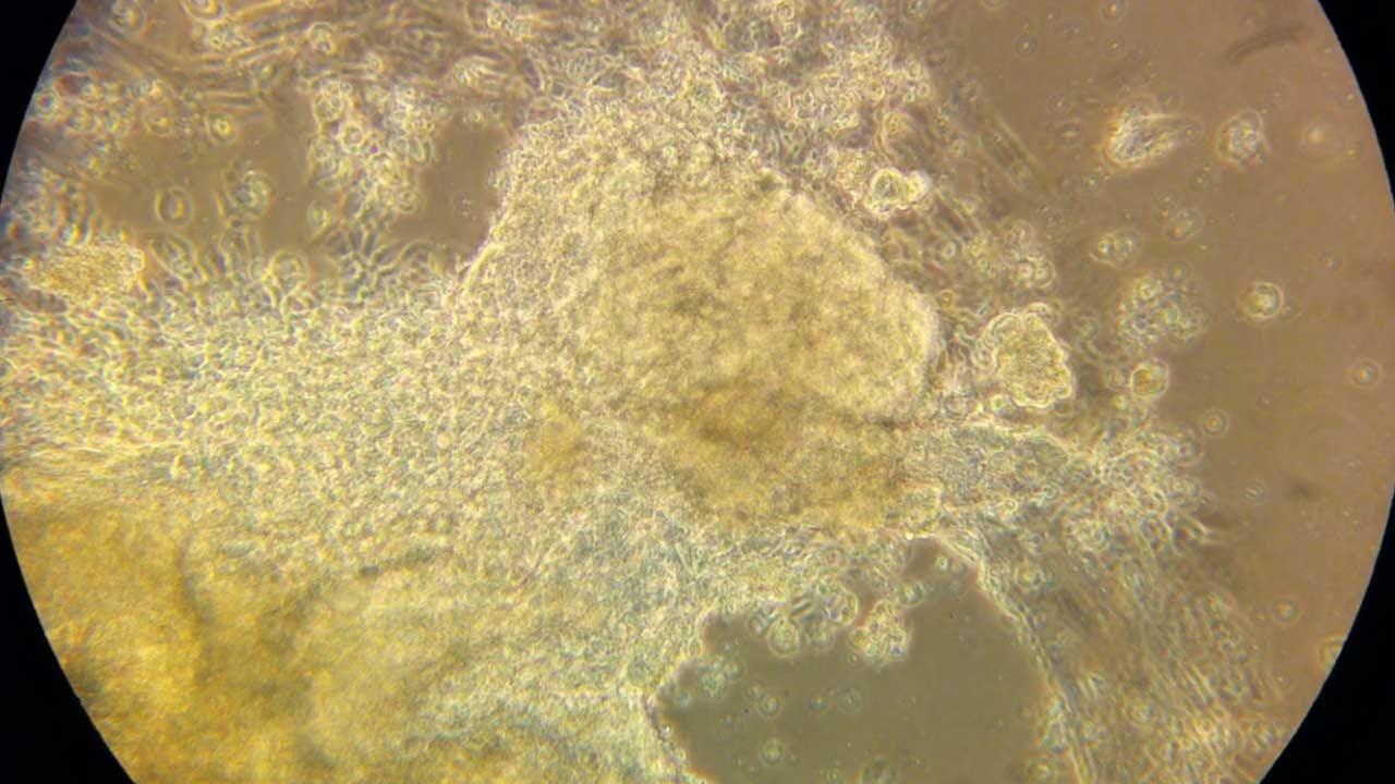
A dish of beating heart cells differentiated from embryonic stem cells
A dish of beating heart cells differentiated from embryonic stem cells. As long as you give it food (fresh culturing media), it will generate a self-paced contraction in a group. Captured by the first prototype of iDu LabCam back in 2011 with a phase-contrast microscope. Credit: Du Cheng, Paula Schanes, Dr. Amy Sprowles.
4. Sperm with hooks
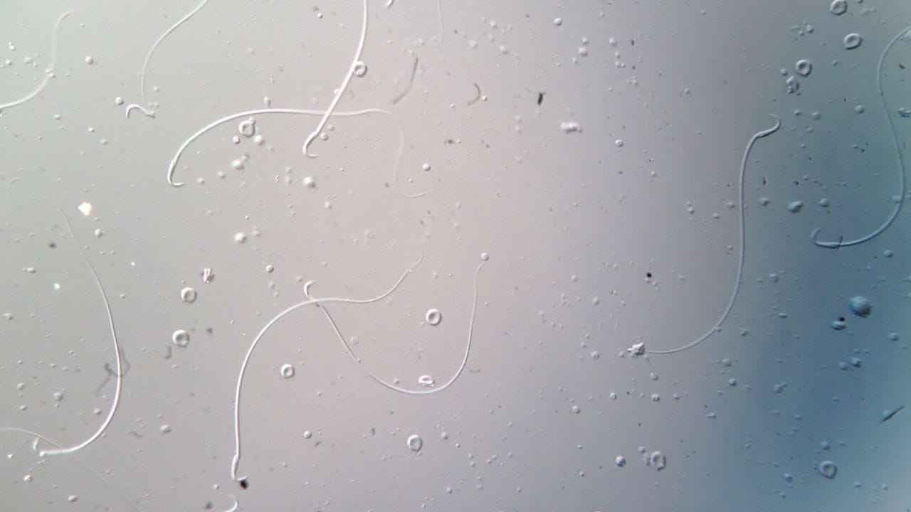
Sperm with hooks!! I guess make sense when we say: hookup. These are not from the movie alien; they are from a rat. Sperm comes in all different shapes and sizes in different species. The differential in sizes and shapes have evolutionary significance. Taken with LabCam on a microscope with Differential Interference Contrast (DIC).
5. Fruit fly mating inside an experimental chamber
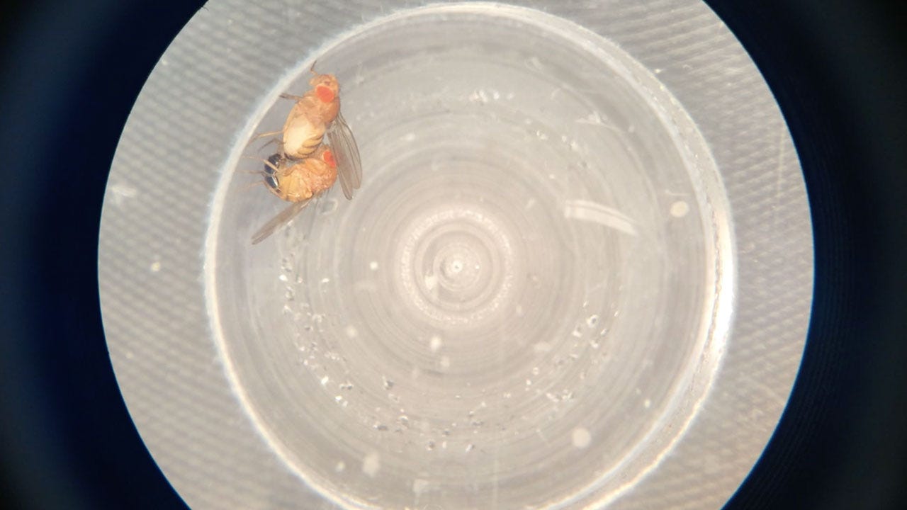
Fruit fly mating inside an experimental chamber
Fruit fly Drosophila melanogaster mating inside an experimental chamber designed to track the detail of their behavior. Fruit fly sing and dance to the female to win her heart over before they mate.
6. Navicula Diatom Time Lapse
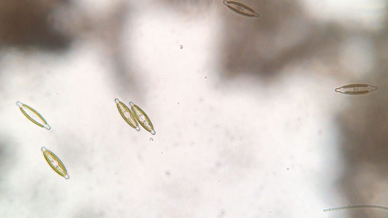
Living in the mud at the bottom of the water garden pond at @brooklynbotanic are many many navicula diatoms. When you look at the pond, you can tell they’re there because they produce tiny air bubbles that sit on the surface of the mud. Diatoms are photosynthetic. Photosynthesis produces oxygen.
Oxygen makes bubbles under water. When you put them under the microscope, you get to see how busy they are. This clip came from Instagram @pondlife_pondlife, taken by Sally Warring with a Leica microscope and LabCam. Sally takes almost all of her videos with LabCam.
7. Cyanobacteria in timelapse
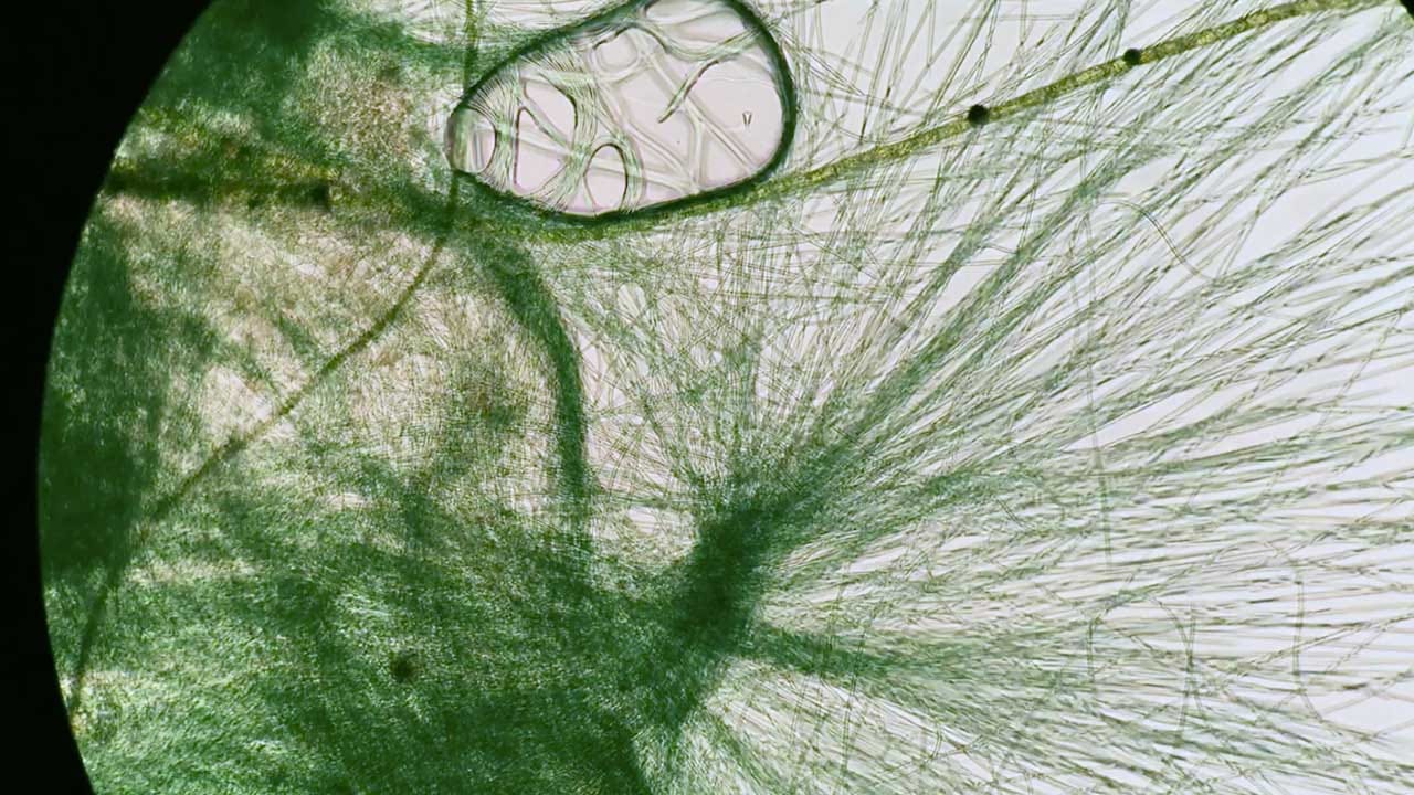
Cyanobacterial motion captured in time lapse. These green guys love to glide around. They sure look good doing it too. This clip came from Instagram pondlife_pondlife, taken by Sally Warring with a Leica microscope and LabCam.
8. LabCam: iDu Optics
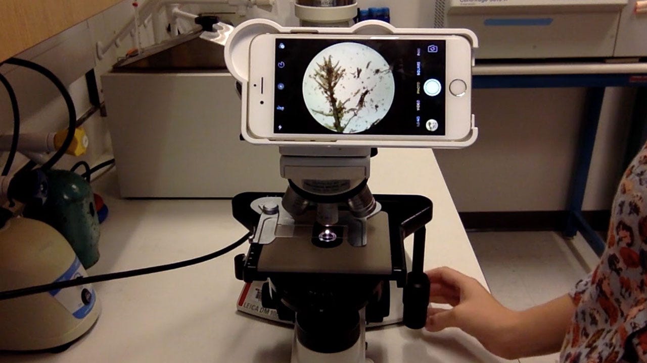
We invented this adapter to help to shoot publication quality picture/video through your microscope with your iPhone with no adjustment needed. We developed this because so many beautiful moments under microscope deserves to be recorded, for an artistic, or scientific purpose. LabCam allows you to turn any microscope into a high quality imaging unit.
About the Author
Du Cheng is currently an MD-PhD student in the Weill Cornell/Sloan-Kettering/Rockefeller U Tri-Institutional MD-PhD program (@weillcornell). Besides learning medicine and doing neuroscience research, he owns a company making LabCam adapter that allows you to take high-quality photos on a microscope with your iPhone (@iLabCam). He is a Paul and Daisy Soros Fellow (@PDSoros) of 2016. For more information on IDu Optics visit: www.iduoptics.com