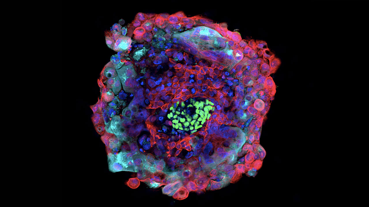
This video depicts Day 6 human blastocyst stage embryo. The cells which would normally become the epiblast and then the entire body are shown here in green. At this stage they are mixed with the red cells which will become the primitive endoderm. The primitive endoderm makes extra-embryonic membranes needed for embryo development, but which do not become part of the body. Even though these cells types are mixed at this stage, the mutually exclusive expression of NANOG (green) and SOX17(red) shows that the cells have already decided whether they will become epiblast or primitive endoderm. The red and the green together make up the inner cell mass (ICM). The inner cell mass is protected and enclosed by a spherical shell of cells: the trophoblast. Cells of the trophoblast (blue nuclei) are linked together geometrically by actin proteins (purple) like a soccer ball. The trophoblast will not contribute to the body of the embryo either, but will go on to form the placenta. The purple trophoblast here is shown pinching into a little nib at one side. This is typical at day 6/7 as the embryo "hatches," erupts out of the zona pellucida, a spherical shell of proteins which surrounds the unfertilized egg, which ensures that only one sperm can enter and fertilize the egg. Sound track: Space Oddity, David Bowie