

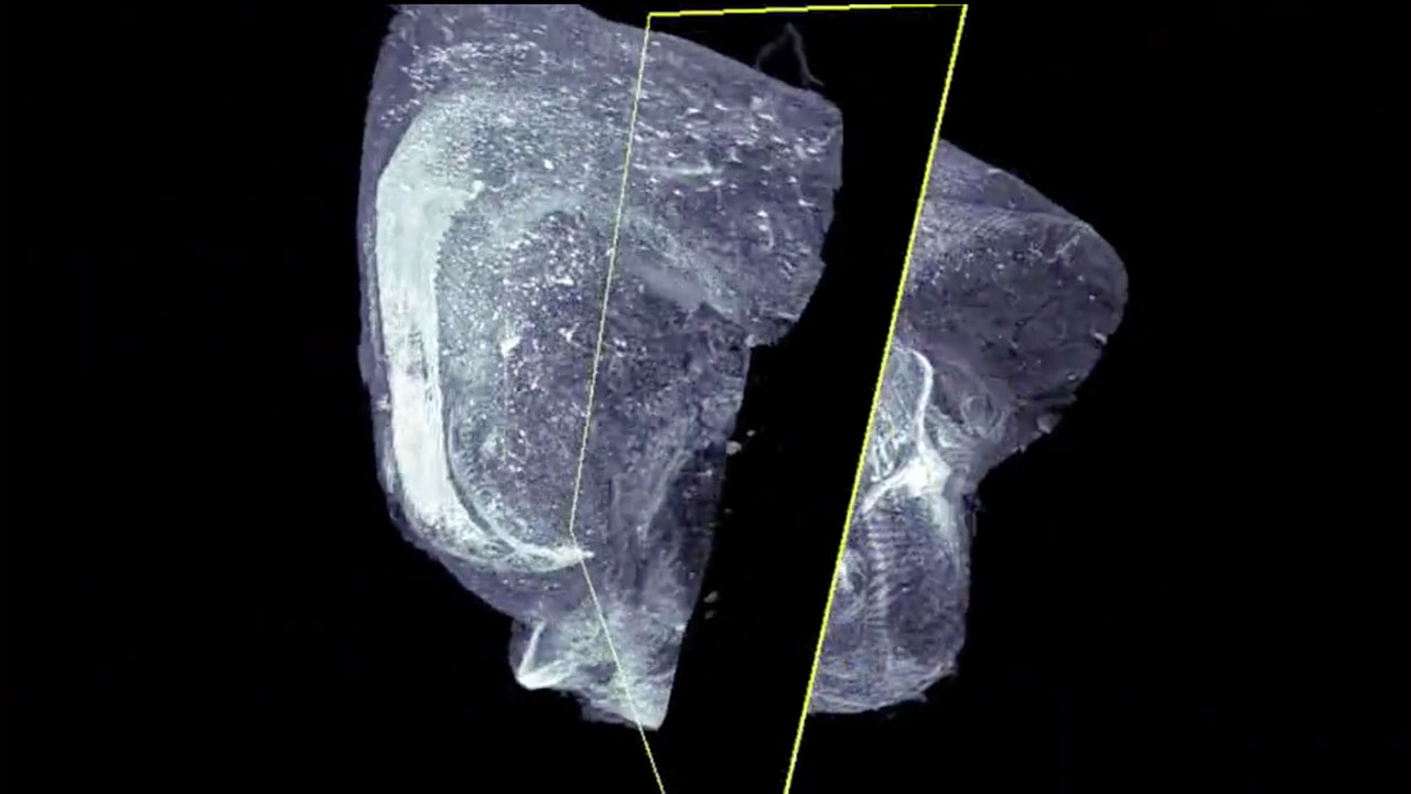
Mouse Embryo Prep for 3D Imaging

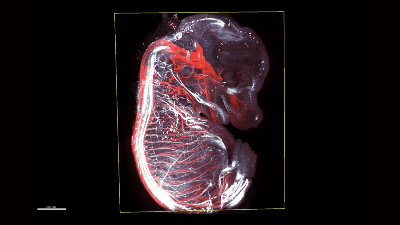
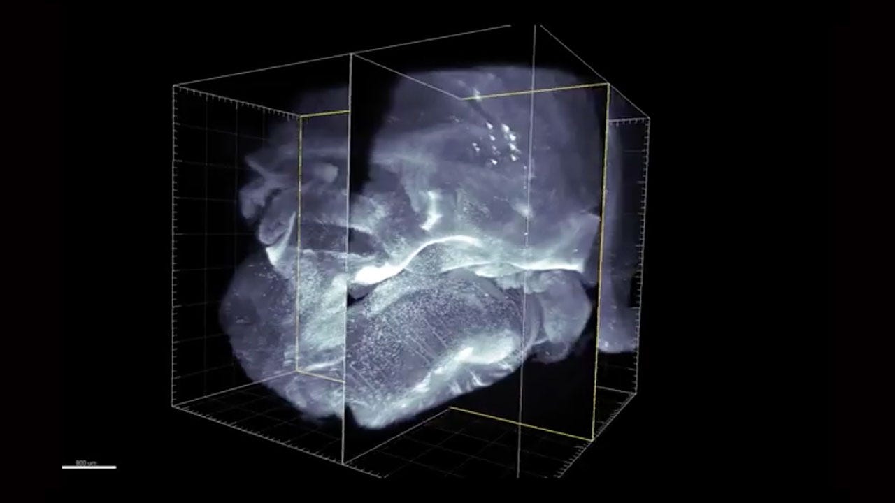
Micro-vasculature of the mouse brain

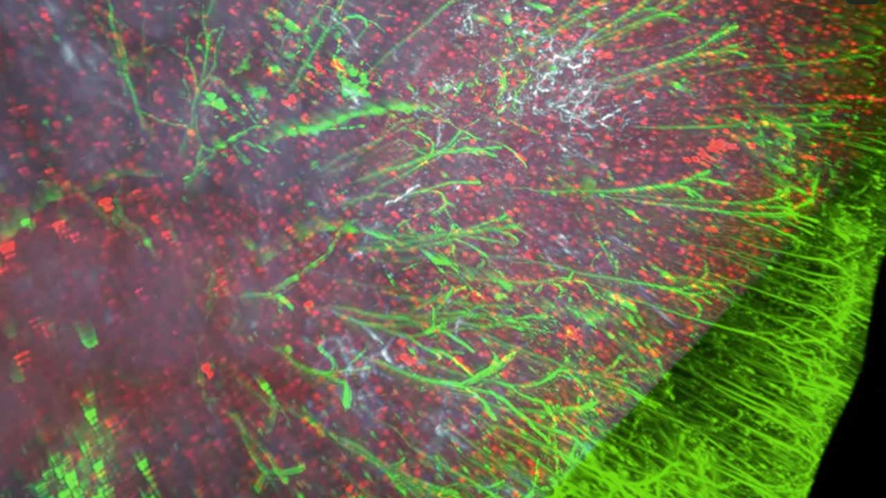
Adrenergic and dopaminergic systems of the adult mouse brain
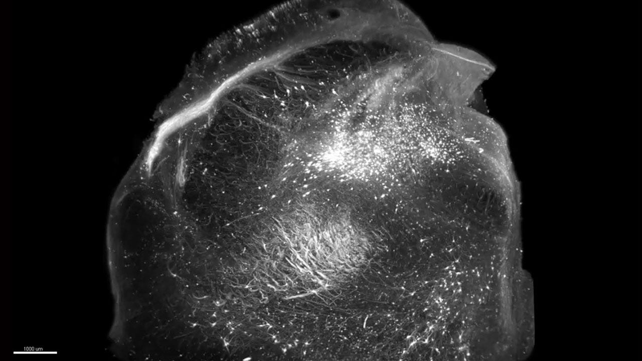
Adult mouse brain showing pyramidal neurons and their projections
Active neurons and Pyramidal neurons in the mouse cortex.

Adrenergic and Dopaminergic systems of the mouse embryo and adult mouse brain

Adrenergic center of the locus coeruleus in a human brain (view of the brainstem)

Cholinergic System of the Adult Mouse Brain
Mouse Embryo at 14 days
Nicolas Renier runs a lab at ICM Brain and Spine Institute in Paris, France, investigating structural plasticity of axons in the brain. He was recently a postdoctoral research fellow at the Rockefeller University, studying how commissural axons in the spinal cord navigate to and across their intermediate targets at the spinal cord midline.
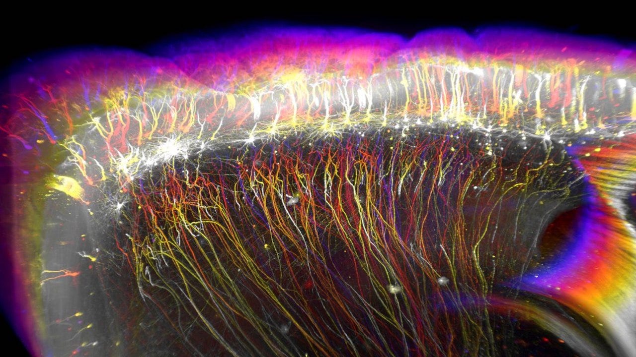
Adult mouse brain showing pyramidal neurons and their projections
Tell us about your research.
I’m interested in finding out how nerves cells get wired together in the brain during development and after, in adulthood. How do we start with lonely neurons, unconnected, and end up with an immensely complex network of connections driving our interactions and experience of the world.

Adrenergic and Dopaminergic systems of the adult mouse brain
More recently, I’ve been interested in how (and also to which extent) those connections can change in the adult brain, for instance when we’re learning new things, or when our brain is affected by an injury or a degenerative disease. The complexity of the problem is such that I had to come up with new ways of visualizing the neurons and their projections. This is how I ended up being interested in imaging brains in 3D.

Active neurons and Pyramidal neurons in the mouse cortex
How do these videos, apart from their aesthetic value, help us understanding axonal navigation and broader questions about brain wiring and function?
This is maybe the most surprising thing: all things considered, we don’t directly work with the videos. We generate the videos or still images occasionally to check the quality of the data (and also because we love to look at them!), but the real scientific work happens after algorithms digest this information for us.
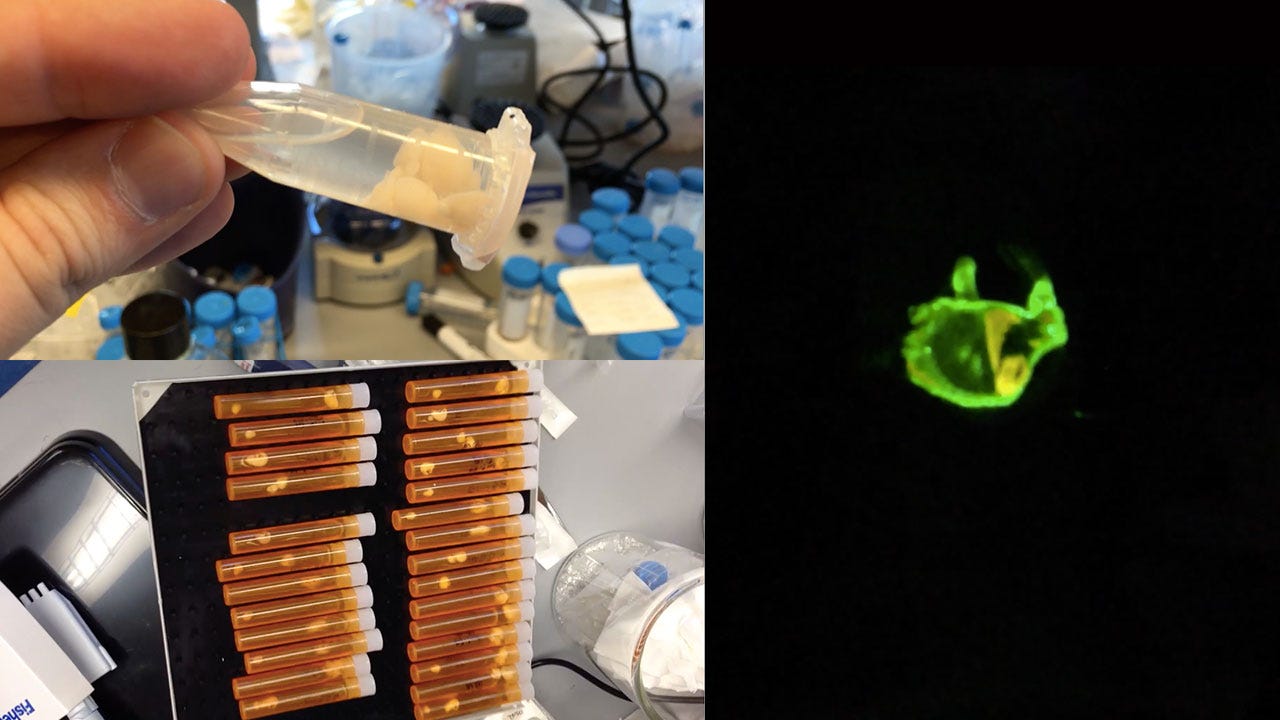
Mouse Embryo Prep for 3D Imaging
The first year of using the 3D imaging technique, we spent a lot of time looking at videos and images of neurons and axons in the brain, but in the end, we couldn’t easily make sense of all this avalanche of information. So we were starting working on algorithms to extract the meaningful information for us, by segmenting the objects and averaging the results of many brains.
In the end, we obtain tables and heat maps, which are not as nice as the real neurons and axons to look at, but provide us the information we’re looking for: where in the brain do the axons of a group of neurons project to? Which neurons are active at a specific point in time?

The advantage of working with intact transparent samples is that we don’t damage anything, as one would do by sectioning, so we can easily segment the axons, the neurons, and many average brains. Thanks to light sheet microscopy, we can acquire each brain in under an hour, which enables us to combine the data from many brains, which was virtually impossible before.
So to answer the original question, the best thing that the volume imaging is bringing us is that ability to streamline the analysis of many samples to try to pull out the meaningful information on axon trajectories and neuron activity, above the natural noise from biological variations.
Describe the techniques used to both do the staining of these whole-mount brains but also tools for creating animations and singular journey through the brain.
In the Tessier-Lavigne lab, my colleague Zhuhao WU and I developed a technique to perform molecular staining on transparent samples we called iDISCO, as a tribute to the original technique we derived it from, 3DISCO. iDISCO stands for “Immunolabeling-enabled 3D Imaging of Solvent Cleared Organs.” The key of the technique is to remove the lipids from the sample to make it permeable to the molecules used to stain the various structures. Most of the time, we’re using antibodies to reveal specific neurons.
After the lipid removal and protein fixation treatment, the brain is like a sponge, and the antibodies can diffuse inside.
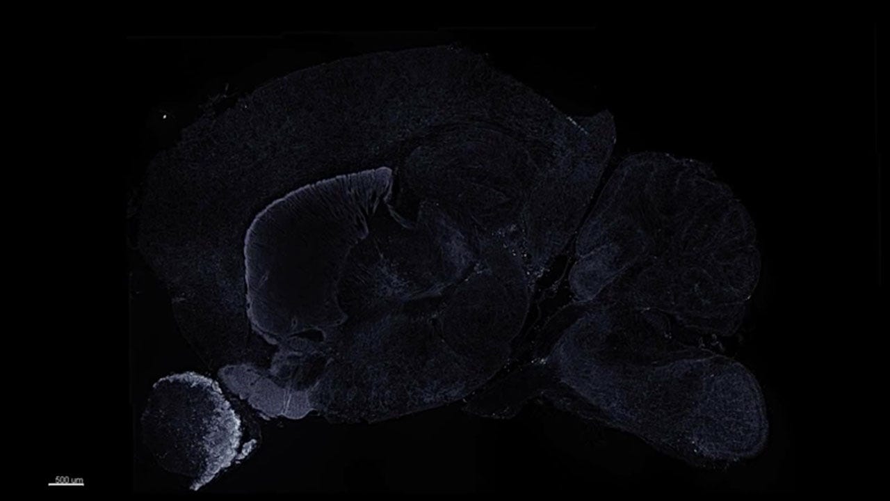
Adrenergic and dopaminergic systems of the adult mouse brain
Once the staining is done, we make the tissue transparent by immersing it in a solvent that has the same optical properties as the brain itself. In physical terms, the speed of light in this solvent happens to be the same as the speed of light going through the brain. So the light can go straight through the brain once immersed in the solvent, making it transparent.
Then, we put the sample under a light sheet microscope. The light sheet microscope scans the sample with a beam of light that has been stretched to look like a sheet of paper (basically a thin plane). This means that by moving this plane through our transparent brain, we can very quickly scan the whole sample.
How do imaging and videos contribute to your scientific work. Apart from its purpose as data, does it have a value for a broader public?
The new type of large-scale, high-resolution 3D data we’re obtaining look like Science Fiction to us scientists. This is because those data are showing us complete structures that we could only previously imagine from sections, or through artistic rendering.
Now we are acquiring a new type of data, straight from the microscope. This is the real biological view of a specimen, and not an interpretation.
I have to admit that maybe part of the lay audience is jaded with this type of 3D images. This may be because 3D renders from artists of biological processes have been around in documentaries or Hollywood movies for a while, so everyone assumes this is how we’re doing Science.
Adrenergic center of the locus in a human brain (view of the brainstem
Those type of 3D renders we’re used to seeing now are the results of years of research to understand the 3D organization of anatomical structures, and they’re an interpretation of the reality. But now, we are acquiring this new type of data, which are coming straight out of the microscope! Those are the real biological view of a specimen, and not an interpretation.
These new visualizations are tremendous for students, to learn anatomy from real microscopic images and not from a textbook rendering. Also, we’re taking advantage of 3D data obtained with this technique and others to create immersive experiences for the public, to travel inside the data and discover the organization of biological objects.
Cholinergic System of the Adult Mouse Brain
“Neurodome” was an initiative in New York to project 3D data of brains and neurons in a planetarium setting, which has been a breathtaking experience for me. Now, with virtual reality headsets, we can engage more people in museums or during Science fairs with those data in a fun way.
Tell us a bit more about yourself.
I have always been fascinated with images and optics. I enjoy photography a lot. To take a step back from the high-tech imaging technique I perform in the lab, I picked up film photography a while back after having shot digital for years. This has been a very refreshing way for me to rediscover this hobby.
I moved recently from New York back to Paris. I feel this is healthy in Science to stay mobile, to change places.
I enjoy greatly picking a roll of film, going out for the day with an old medium format camera, and doing the processing and printing later at home. This way of doing photography constrains me within the limitation of the film and camera I picked. This is a more satisfying experience than shooting digital, where you can alter more things in post.
This is a bit similar to an experiment in the lab: you’re not going to get everything you want from your sample and your microscope, and you have to work with those limitations in mind.
I moved recently from New York back to Paris. I feel this is healthy in Science to stay mobile, to change places.
This brings a lot of fresh air into your way of thinking, but more importantly you meet new people and can start new things from those interactions. I benefited tremendously from the scientific environment at Rockefeller University, with colleagues such as Zhuhao Wu, Christoph Kirst and others who were key in making this project successful. So I’m looking forward to the new things Paris will bring, which is exciting and stressful at the same time!
Interview conducted by Alexis Gambis, Executive Director of Imagine Science Films