Evolution is a unifying paradigm with important implications in virtually all areas of biology. Indeed, all characters that are studied in biology, from morphological or behavioural traits to the immune system or the fine regulatory mechanisms of gene expression are the products of biological evolution. Evolutionary concepts are pertinent not only to that large diversity of possible applications across disciplines, but also to the massive realm of diversity across living beings: from natural/artificial clones to the major kingdoms made of approximately 50 millions of extant species, all living beings are connected through pedigrees and the phylogenetic tree of life.
 A male panther chameleon (Furcifer pardalis) in Madagascar. Credit: Michel C Milinkovitch
Michel C. Milinkovitch and his team recently published a Nature article entitled "A living mesoscopic cellular automaton made of skin scales" (view only version). For this occasion, we sat down with him to learn more about his research on skin colour patterns in lizards and the importance of photography and filmmaking to capture these dynamic processes.
The three pictures of panther chameleons illustrates the very large colour variation that exists in Furcifer pardalis. Our genetics analyses actually show that what was thought to be a single species should be divided in about 11 species.
Interview conducted by Alexis Gambis, Executive Director of Imagine Science Films

A close up on the skin of an ocellated lizard (Timon lepidus) whose pattern is produced by a hexagonal cellular automaton, in which the skin scales are the discrete units. Credit: Michel C. Milinkovitch
Our work is targeted to biologists who wish to understand the importance of physical processes in biological systems as well as to physicists interested to learn more about the living world as complex physical systems. More generally, our audience is everyone interested into the complexity, diversity and beauty of the living world.
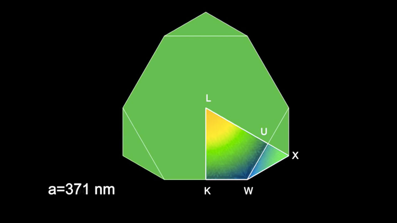
We study colours and colour patterns (www.lanevol.org), so images and videos are paramount to convey the results of our research. These movies show the process at work at the nanoscale level (movie S5) and how it translates at the cellular level (movie S4) and at the macroscopic in-vivo scale (Movies S1 and S2).

A second male panther chameleon (Furcifer pardalis) in Madagascar. Credit: Michel C Milinkovitch
The three pictures of panther chameleons illustrates the very large colour variation that exists in Furcifer pardalis. Our genetics analyses (http://onlinelibrary.wiley.com/doi/10.1111/mec.13241/full ) actually show that what was thought to be a single species should be divided in about 11 species.
Techniques in the featured videos
For S1, S2 and S3, we use a simple high-resolution video camera.
For S4: a simple high-resolution camera fixed on a microscope.
For S5: a numerical simulation performed 'in silico' (on a computer)
These videos were made in my lab by Jérémie Teyssier (a physicist), Suzanne Saenko (a biologist) and myself (biophysicist). They generated a lot of interest both in the scientific community and the media (national Geographic, BBC, PBS, multiple other magasines, newspapers, TV programs, blogs, YouTube channels etc). Note that the focus should not be us (as scientists) but the chameleons (our object of research): they are magnificent and allow us to appreciate the beauty and complexity of living systems.
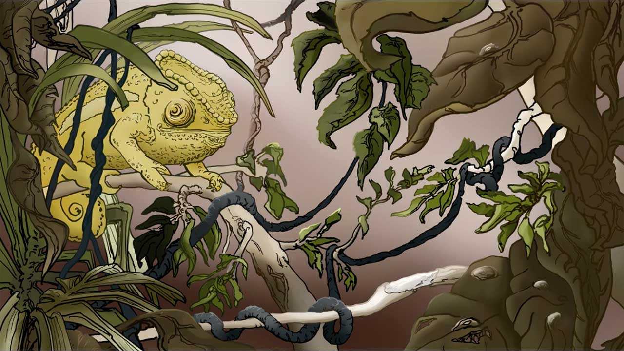
Check also the animation video made by Maya Hartmeier during her graduation project at the ZHdK (Zurich University of Arts) to explain our work on chameleon colour change.
Maya built some cool animations to explain how chameleons play with the light interference phenomenon to shift colour through the active tuning of a lattice of guanine nanocrystals within dermal iridophores. Additional information is available on the ‘Chameleon Colour Change’ page and in the original article

The third panther chameleon indiviudal. Credit: Michel C Milinkovitch
About Author
Michel Milinkovitch is Full Professor in the Department of Genetics & Evolution at the University of Geneva (Switzerland). He is also a member of the Institute of Genetics and Genomics in Geneva (iGE3) since its foundation (2011) and a Group leader of the Swiss Institute of Bioinformatics (SIB) since 2014. As an evolutionary geneticist, he contributed significantly to quantitative analysis & modelling in Molecular Phylogenomics and Applied Evolutionary Genetics. He has developed concepts, analytical tools, and algorithms / models for multiple sequence alignments, phylogeny inference and haplotypic network building. His recent focus is on Evolutionary Developmental Genetics (Evo-Devo) and the Physics of Biology. He has published over 100 papers in international peer-reviewed journals for a cumulate impact factor of over 900. He has also published 7 book chapters. He has given over 160 talks all around the world. He has been reviewer for over 30 peer-reviewed international joufrnals and for academic promotions, grant proposals, and PhD committees in Europe and the USA. He has served, and is still serving, on the Editorial Board of scientific journals and has supervised more than 15 PhD theses. He has served on the ERC (European Research Council) evaluation panel. Michel Milinkovitch obtained several prizes and awards both for his academic and popularisation work. He is co-founder of the spin-off Delphi Genetics.
All our research is highly collaborative. Jeremie Teyssier is a fantastic post-doc who was trained as a physicist but he is also very much interested in photonic effects in the living world. Suzanne Saenko has been working as a developmental biologist in butterfly colour formation during her PhD and moved to chameleons and other lizards in my lab. We are now also collaborating with first-class biochemists and cell biologists such as Marcos Gonzalez-Gaitan to understand how cells can fabricate nanocrystals.